
“A Simple working human heart model with electronics for visualization of arrhythmia.
The main objective of this project is to teach students how the human heart functions.
The heart has 4 chambers and 4 valves: Right Atrium, Left Atrium, Right Ventricle, and Left Ventricle. Tricuspid Valve, Mitral Valve, Aortic Valve, Pulmonic Valve. - The valves prevent the backflow of blood
The human heart has two types of muscle cells for functioning. 1. Contractile cells: Helps in contraction and relaxation of the heart2. Conducting cells: They carry the electricity throughout the heart to contractile cells.
BasicPhysiology:
CirculatorySystem:
The right side of the heart (RightAtrium) receives the deoxygenated blood from the Superior and Inferior vena cava and sends it to the Right Ventricle via the Tricuspid valve. From the Right ventricle, the blood ejects out to the lungs for purification via the Pulmonic valve.
The left side of the heart (Left Atrium) receives the oxygenated blood from the lungs through 4 pulmonary veins and sends it to the Left Ventricle via the mitral valve. From the Left Ventricle, blood is ejected out to the whole body for circulation via the aortic valve.
Conduction System:
SA Node: Natural pacemaker of the heart. Electrical Impulses begin here. AV Node: It lies in between the Atria and ventricles. The impulse from the SA node reaches the AV node. The impulse delays a bit for ventricular filling. Bundle of HIS and Branches: The impulse from the AV node reaches His bundle and bifurcates into Left and Right BundleBranches.Purkinje Network: The Purkinje network layers up both the ventricles and impulses will make the ventricles to contract and eject the blood out.
Understanding Arrhythmias:
Any deviation from normal rhythm is termed arrhythmia.
SA node - P wave, AV node - PR Interval, Bundle Branches and purkinje network - qRS complex, Ventricle Relaxation - T wave
Normal Sinus Rhythm: SA -> AV -> Bundle branches -> Purkinje networks. Normal intrinsic rate 60-100 bpm. Tachycardia > 100 bpm, Bradycardia < 60 bpm.
Junctional Rhythm: The SA node is blocked or delayed. Intrinsic rate: 40-60 bpm. AV -> Bundle branches -> Purkinje networks. Accelerated Junctional: 60-100 bpm Junctional tachycardia > 100 BPM
Ventricular Rhythm: Both SA and AV node stops producing impulses. ventricles take charge by producing impulses with an intrinsic rate of 20-40 bpm. Accelerated Ventricular Rhythm: 40-100 bpm, Ventricular tachycardia >100 bpm.
First Degree AV Block: When the SA node fires the impulse but delays more than 120 s in the AV node.
Second Degree AV Block:Type 1: Some impulses get dropped off without reaching the ventricles. The PR interval will be changing constantly. Type 2: Some impulse dropped without conducting the ventricles with a constant PR interval.
Third Degree AV Block: The SA node generates the impulses randomly and sometimes it will get conducted. the ventricular rhythm overrides the situation. Both Impulses fire randomly without sync.
Pulseless Electrical Activity: The heart chambers will not contract but the electrical activity will be ongoing. Its an medical emergency.
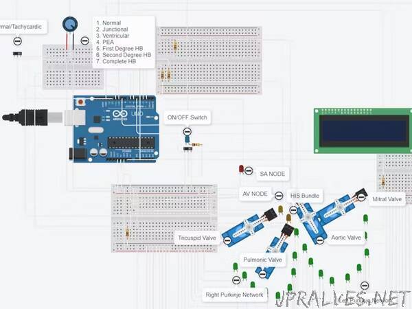
Proposed System: A slideswitch switch to turn on/off the simulator. The potentiometer to tune in the rhythm. Other slideswitch to toggle tachycardia on and off. The conduction system network is shown with LEDs. The 4 servo motors acts as 4 heart valves. A LCD display screen to display the selection.
I hope with the help of this project, students will be able to understand the mechanism of basic arrhythmia.
Future Advancements: The existing system only has few rhythms, The aim is to simulate other complicated arrhythmias in this model.”