
“ETH researchers have managed to take three-dimensional pictures of single nanoparticles using extremely short and strong X-ray pulses. In the future this technique could even be used to make 3D-movies of dynamical processes at the nanoscale.
X-ray diffraction has been used for more than a hundred years to understand the structure of crystals or proteins – for instance, in 1952 the well-known double helix structure of the DNA that carries genetic information was discovered in this way. In this technique, the object under investigation is bombarded with short-wavelength X-ray beams. The diffracted beams then interfere and thus create characteristic diffraction patterns from which one can gain information about the shape of the object.
For several years now it has been possible to study even single nanoparticles in this way, using very short and extremely intense X-ray pulses. However, this typically only yields a two-dimensional image of the particle. A team of researchers led by ETH professor Daniela Rupp, together with colleagues at the universities of Rostock and Freiburg, the TU Berlin and DESY in Hamburg, have now found a way to also calculate the three-dimensional structure from a single diffraction pattern, so that one can “look” at the particle from all directions. In the future it should even be possible to make 3D-movies of the dynamics of nanostructures in this way. The results of this research have recently been published in the scientific journal Science Advances.
Daniela Rupp has been assistant professor at ETH Zurich since 2019, where she leads the research group “Nanostructures and ultra-fast X-ray science”. Together with her team she tries to better understand the interaction between very intense X-ray pulses and matter. As a model system they use nanoparticles, which they also investigate at the Paul Scherrer Institute. “For the future there are great opportunities at the new Maloja instrument, on which we were the first user group to external pagemake measurements at the beginning of last yearcall_made. Right now our team there is activating the attosecond mode, with which we can even observe the dynamics of electrons,” says Rupp.
A deeper view into dynamical processes
The recently published work is an important step towards that future, as postdoctoral researcher Alessandro Colombo explains: “With this work, we open a window on studies of dynamical processes of the extremely small particles in the femtosecond regime.” The problem with X-ray diffraction using very intense pulses is that the objects under investigation evaporate immediately after the bombardment – “diffract and destroy” in the researchers’ jargon. Since this means that only a single snapshot of the nanoparticle can be made, of course one would like to obtain as much information as possible from it. To compute more than a 2D image from the diffraction pattern, up to now one had to impose on the computer algorithm some strongly limiting assumptions on the shape of the nanoparticle, for instance its symmetry. However, in this way any fine detail of the particle that deviates from those assumptions remains hidden. Moreover, with those algorithms many adjustments had to be made by hand.
Improved algorithm
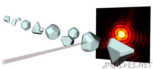
“This is where our new method comes in”, says Rupp: “With our new algorithm, which uses a very efficient simulation method and a clever optimization strategy, we can automatically produce 3D images of the nanoparticle without having to impose specific requirements. This allows us to see even tiny irregularities, which can arise from the growth process of the particle.” To achieve 3D resolution, the researchers at ETH do not just use that part of the diffraction pattern which is diffracted by a small angle of a few degrees, as has been customary up to now, but also the wide-angle part of 30 degrees or more. This means, of course, that the amount of information to be retrieved increases enormously, but the improved algorithm can cope even with that.
In this way, from the diffraction patterns of single silver nanoparticles 70 nanometers in size that are bombarded with X-ray pulses lasting around 100 femtoseconds, Rupp’s team can now calculate 3D pictures that show the particles from different angles.
Snapshots in free flight
“Up to now we were missing that third dimension”, says Rupp,”but now we can investigate many processes either for the first time or with unprecedented precision, for instance, how nanoparticles melt in a few picoseconds or how nanorods accumulate to form larger objects. “ The crucial point is that the snapshots can be taken in free flight in vacuum, without having to fix the nanoparticles on a surface, as is done in electron microscopy. Moreover, many kinds of particles cannot even be put on a surface because they are too fragile or short-lived. But even those samples that can be studied with an electron microscope are considerably influenced by their interaction with the surface. In free flight, on the other hand, melting or aggregation processes can be studied without any disturbance.
Reference
Colombo A et.al.: Three-dimensional femtosecond snapshots of isolated faceted nanostructures. Science Advances, 22. February 2023, doi: external page10.1126/sciadv.ade5839”