“When a person experiences a happy or sad mood, which brain cells are active?
To answer that question, scientists need to understand how individual brain cells contribute to a larger network of brain activity, and what role each cell plays in shaping behavior and overall health. Until now, it’s been difficult to get a clear view into how brain cells in living animals behave over extended periods of time.
But Jia Liu’s group at the Harvard John A. Paulson School of Engineering and Applied Sciences (SEAS) has developed an electronic implant that collected detailed information about brain activity from a single cell of interest for more than a year. Their findings, based on research in mice, are reported in Nature Neuroscience.
“This research solves a fundamental issue – the challenge of creating a brain-electronic interface that does not disturb brain function or degrade over time,” says Liu, who is an assistant professor of bioengineering at SEAS, where he leads a lab dedicated to bioelectronics.
Neuroscientists have long sought better tools to study different cells in the brain, including neurons (which transmit electrical and chemical messages) and microglia (immune cells responsible for maintaining brain health).
“A single neuron is very small — only 10 to 100 micrometers — and when it fires, its action potential (the spike in electrical activity) only lasts about two milliseconds,” Liu says.
Certain techniques can detect brain activity from specific cells of interest for short-lived experiments in small areas of the brain, either in tissue recently removed from animals or by using probes or optogenetic techniques to capture activity in situ.
But these conditions are not “true to life” and they don’t provide detailed enough information about electrical activity in individual cells to understand how activity changes with age and other life experiences, Liu says. “Behaviors, memories, and disease all build up over the course of days, weeks, months, and years.”
Much of the difficulty to date, he says, has been due to a mismatch in mechanical properties between living brain tissue and electronic recording devices. This has prevented long-term, precise recording of how neurons and microglia behave over time.
“The brain is very soft, like the texture of tofu or pudding. In contrast, electronics are rigid. Any small movement of the brain can cause conventional sensors to drift and move in living brain tissue. That mismatch in structure can cause cells around the implantation site to degrade.”
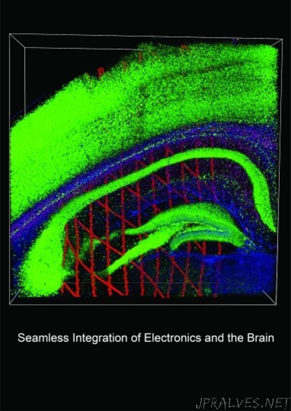
To circumvent the problem, Liu’s team, which specializes in engineering nanoelectronics or “cyborgs” to bridge the gap between living tissue and electronics, developed an implantable device and minimally invasive technique for delivering it safely into the brain. The mesh-like, flexible nanoelectronic sensor is designed to be inserted into brain tissue using a water-soluble polymer “shuttle.” Prior to implantation, the device and its delivery shuttle are connected lithographically. Once the implant is in the brain, a simple saline solution is applied to dissolve the shuttle, leaving only the mesh electronic sensor behind.
In mouse studies, when Liu’s team implanted their nanoelectronic sensors into multiple areas of the brain, the implantation process and presence of the sensors resulted in minimal disturbance to brain tissue. Then, targeting single neurons for analysis, they used the devices to record the electrical activity of those same cells over the course of the mice’s adult lives.
“Even after one year, we didn’t see any degradation of the individual neurons or proliferation of the microglia we were interested in recording with the devices,” Liu says. “There’s no other technology out there that can track single-cell action potential from the same cells in active animals over the course of a few months and a year.”
Looking ahead, Liu plans to further develop the technique so that brain activity can be transmitted in real time from the biological neural network to an artificial neural network in a computer for analysis. And, he wants to explore how the mesh nanoelectronic sensors can be used to study phenomena such as “neural representation.”
“When you watch a movie or see a car drive down the road, your brain generates electrical activity to represent those images,” he says. During that process of neural representation, the brain encodes sensory information and thoughts into a model of external stimuli.
Liu says that, for example, moods are influenced by neural representation, and he’s especially interested in studying how changes of neural representations and brain states impact mood fluctuations over time.
“Maybe one day it’s cold and gray outside, and you feel unhappy and in a bad mood. Another day, it’s sunny and you’re on the beach and you’re in a great mood. How those representations change in the brain cannot be studied by current technology because we haven’t been able to stably track activity from the same neuron,” he says. “This research completely overcomes that limitation. It’s the beginning of a new era of neuroscience.”
An ultimate goal of Liu’s research is to develop diagnostic and therapeutic methods for neurological, cardiovascular and developmental diseases.
Additional authors are Siyuan Zhao, Xin Tang, Weiwen Tian, Sebastian Partarrieu, Ren Liu, Hao Shen, Jaeyong Lee, Shiqi Guo, and Zuwan Lin.
This work was supported by SEAS and the Harvard University Faculty of Arts and Sciences Dean’s Competitive Fund for Promising Scholarship.”
