
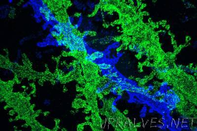
“MIT researchers have developed a way to make extremely high-resolution images of tissue samples, at a fraction of the cost of other techniques that offer similar resolution. The new technique relies on expanding tissue before imaging it with a conventional light microscope. Two years ago, the MIT team showed that it was possible to expand tissue volumes 100-fold, resulting in an image resolution of about 60 nanometers. Now, the researchers have shown that expanding the tissue a second time before imaging can boost the resolution to about 25 nanometers. This level of resolution allows scientists to see, for example, the proteins that cluster together in complex patterns at brain synapses, helping neurons to communicate with each other. It could also help researchers to map neural circuits, says Ed Boyden, an associate professor of biological engineering and brain and cognitive sciences at MIT.”