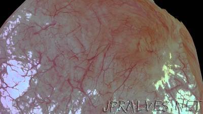
“The way doctors examine the bladder for tumors or stones is like exploring the contours of a cave with a flashlight. Using cameras attached to long, flexible instruments called endoscopes, they find that it’s sometimes difficult to orient the location of masses within the bladder’s blood vessel-lined walls. This could change with a new computer vision technique developed by Stanford researchers that creates three-dimensional bladder reconstructions out of the endoscope’s otherwise fleeting images. With this fusion of medicine and engineering, doctors could develop organ maps, better prepare for operations and detect early cancer recurrences.”
Related Content
Related Posts:
- Exploring the ultrasmall and ultrafast through advances in attosecond science
- A replacement for traditional motors could enhance next-gen robots
- New high-speed microscale 3D printing technique
- Researchers show an old law still holds for quirky quantum materials
- Groundbreaking study shows defects spreading through diamond faster than the speed of sound
- ‘Computer vision’ reveals unprecedented physical and chemical details of how a lithium-ion battery works
- Unlocking the mysteries of freezing in supercooled water droplets
- A molecular additive enhances next-gen LEDs – but shortens their lifespans
- Researchers show how to increase X-ray laser brightness and power using a crystal cavity and diamond mirrors
- New nontoxic powder uses sunlight to quickly disinfect contaminated drinking water