
“A groundbreaking microscopy technique that makes it possible to study biomolecules, nature’s smallest biological building blocks, in a new, more efficient way. This is presented by researchers from Chalmers and University of Gothenburg, using so-called nanochannels to study the molecules.
“This new microscopy method opens new possibilities towards studying small biomolecules. This will allow us to gain new insights into the biological function of cells and tissues,” says Giovanni Volpe, Professor at the Department of Physics at the University of Gothenburg.
Biomolecules are both small and elusive, but vital since they are the building blocks of everything living. In order to get them to reveal their secrets using optical microscopy, researchers currently need to either mark them with a fluorescent label or attach them to a surface.
“With current methods you can never quite be sure that the labelling or the surface to which the molecule is attached does not affect the molecule’s properties. With the aid of our technology, which does not require anything like that, it shows its completely natural silhouette, or optical signature, which means that we can analyse the molecule just as it is,” says research leader Christoph Langhammer, professor at the Department of Physics at Chalmers. He has developed the new method together with researchers in both physics and biology at Chalmers and the University of Gothenburg.
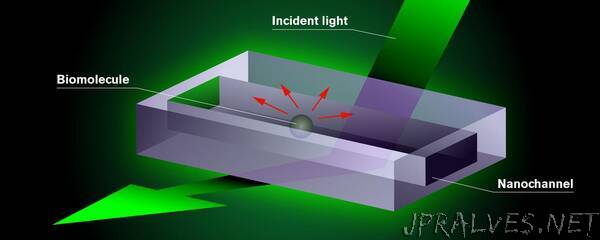
The unique microscopy method is based on those molecules or particles that the researchers want to study being flushed through a chip containing tiny nano-sized tubes, known as nanochannels. A test fluid is added to the chip which is then illuminated with visible light. The interaction that then occurs between the light, the molecule and the small fluid-filled channels makes the molecule inside show up as a dark shadow and it can be seen on the screen connected to the microscope. By studying it, researchers can not only see but also determine the mass and size of the biomolecule, and obtain indirect information about its shape – something that was not previously possible with a single technique.
“The next step will be to push the limit of resolution of the method down towards ever smaller molecules. In this context, the use of the AI-enhanced microscopy software DeepTrack, which we have developed, analysis will prove critical,” says Giovanni Volpe.”