
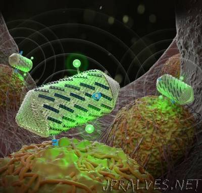
“Ultrasound imaging is used around the world to help visualize developing babies and diagnose disease. Sound waves bounce off the tissues, revealing their different densities and shapes. The next step in ultrasound technology is to image not just anatomy, but specific cells and molecules deeper in the body, such as those associated with tumors or bacteria in our gut. A new study from Caltech outlines how protein engineering techniques might help achieve this milestone. The researchers engineered protein-shelled nanostructures called gas vesicles—which reflect sound waves—to exhibit new properties useful for ultrasound technologies. In the future, these gas vesicles could be administered to a patient to visualize tissues of interest. The modified gas vesicles were shown to: give off more distinct signals, making them easier to image; target specific cell types; and help create color ultrasound images.”